
Omics Data
Single-Cell RNA-sequencing to profile T cells across organs
Mapping Where and How Immune Cells Act in Different Organs
Overview
We analyzed a publicly available single-cell RNA sequencing data of mouse γδ T cells collected across multiple tissues (liver, lung, small intestine, large intestine) from the study: Multimodal profiling reveals site-specific adaptation and tissue residency hallmarks of γδ T cells across organs in mice (du Halgouet, A. et al. Nat Immunol 25, 343–356 (2024))
Research objective: How do γδ T cell states differ across tissues, and which immune programs dominate in each environment?
Using Drylab, the dataset was processed end-to-end — from raw data to annotated cell states and tissue mapping — in a single workflow.
Key findings:
Cytotoxic intraepithelial lymphocytes (IELs) dominate intestinal tissues
IL-17-producing γδ T cells (gdT17) are enriched in the lung
Activated/stress-associated states accumulate in the liver
gdT17 represents a transcriptionally distinct immune program, separating clearly from other cell states
Drylab also identified and excluded a low-quality experimental batch with poor labeling signal, ensuring that all conclusions were based on high-confidence data.
Scientific Deep Dive
Data Processing and Quality Control
Single-cell multiome data (scRNA-seq with HTO labeling) from γδ T cells were processed using an end-to-end workflow including quality control, normalization, highly variable gene selection, PCA, and graph-based clustering.
HTO-based demultiplexing was performed to assign tissue-of-origin. One batch (DEC) exhibited low-quality, ambient-dominated HTO signal and was excluded from downstream analysis. All subsequent tissue-resolved analyses were conducted on the OCT batch, comprising 10,125 high-confidence cells across liver, lung, small intestine, and large intestine
Clustering and Cell State Annotation
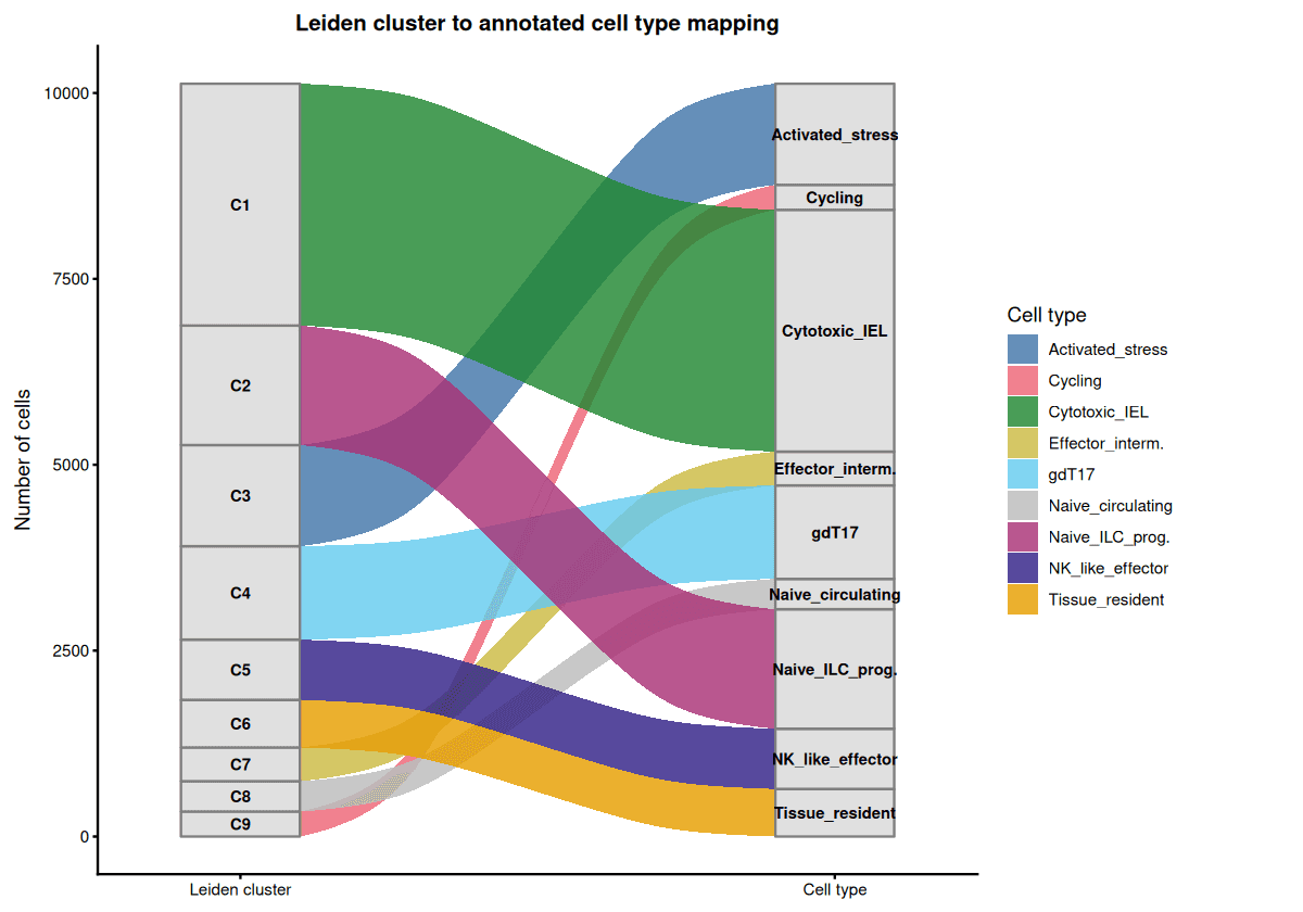
Leiden clustering (resolution = 0.5) identified 9 transcriptionally distinct clusters, each mapping one-to-one to biologically defined γδ T cell states.
Cell type distribution (post-contaminant cleanup, n = 10,125 cells from OCT batch): Cytotoxic_IEL is the dominant population (C1, ~32%), followed by Naive_ILC_progenitor (C2, ~16%) and gdT17 (C4, ~12%)
Differential expression analysis (Wilcoxon rank-sum test) was used for annotation.
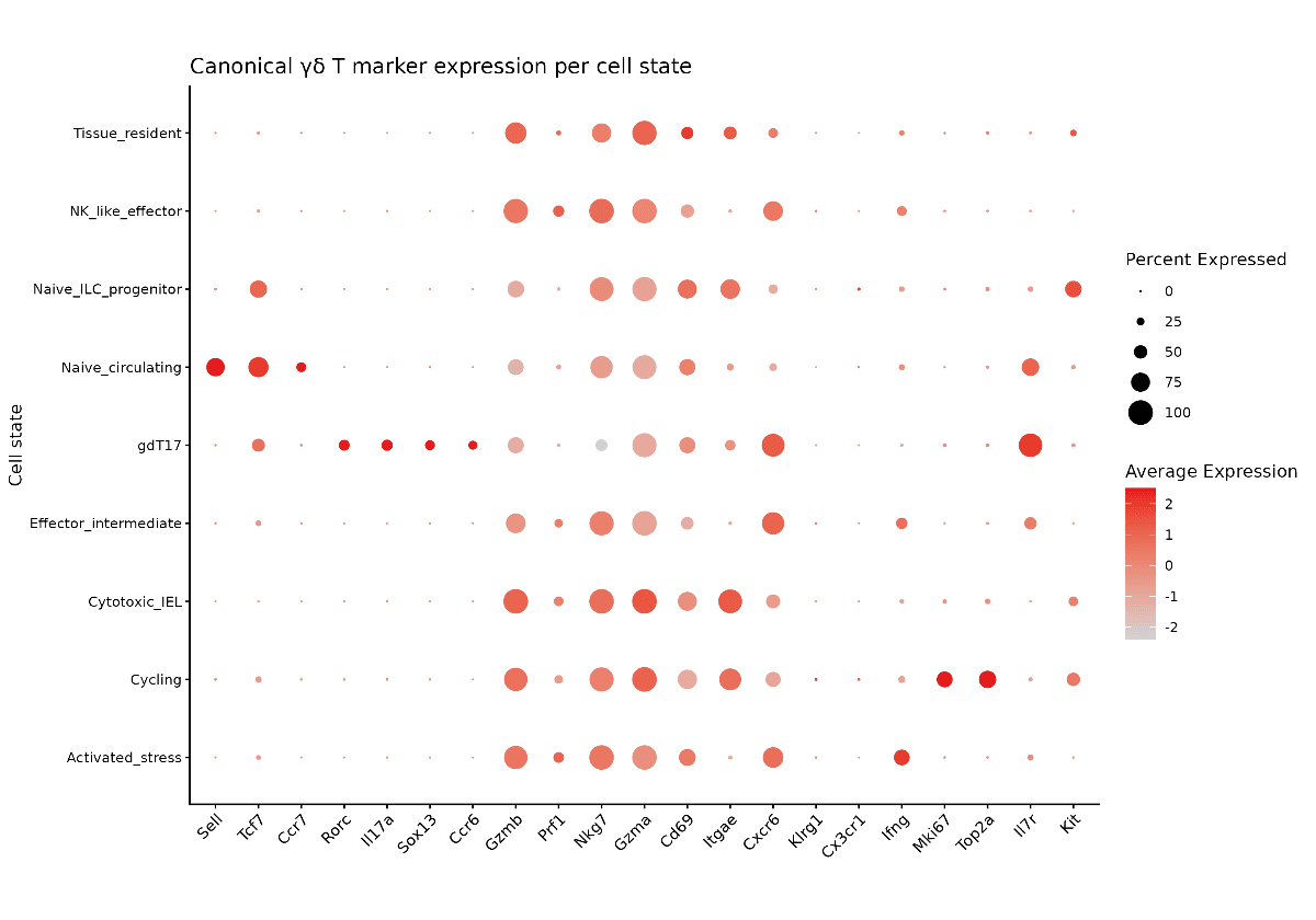
Canonical marker validation confirmed expected transcriptional programs:
Naive states: Sell, Tcf7
gdT17: Rorc, Il17a
Cytotoxic populations: Gzmb, Gzma, Prf1
Cycling cells: Mki67, Top2a
Tissue-Resolved Immune Architecture
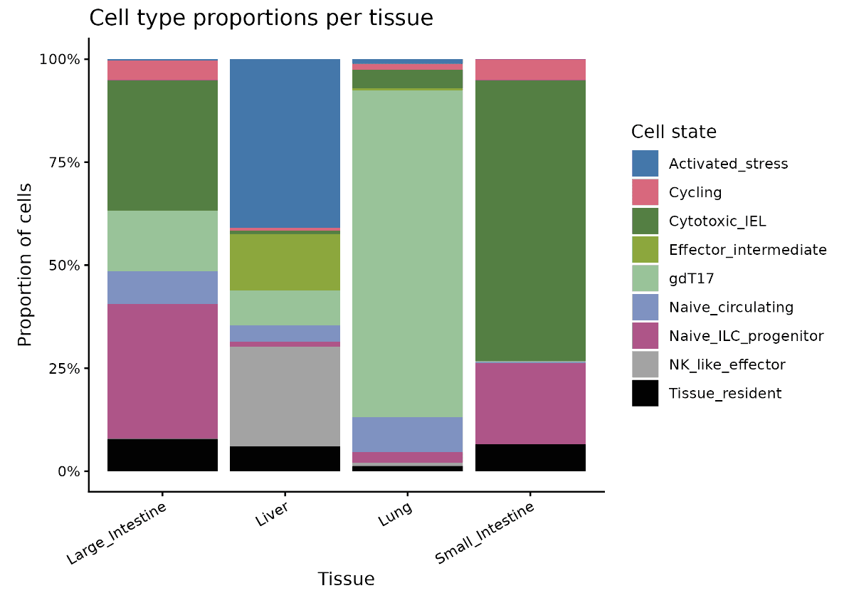
Integration of HTO labels enabled mapping of cell states to tissue compartments. Distinct spatial enrichment patterns were observed:
Cytotoxic intraepithelial lymphocytes (IELs) were enriched in intestinal tissues (SI, LI)
gdT17 cells were preferentially localized to the lung
Activated/stress-associated states accumulated in the liver
These patterns are consistent with known tissue-specific immune functions and support the biological validity of the inferred states.
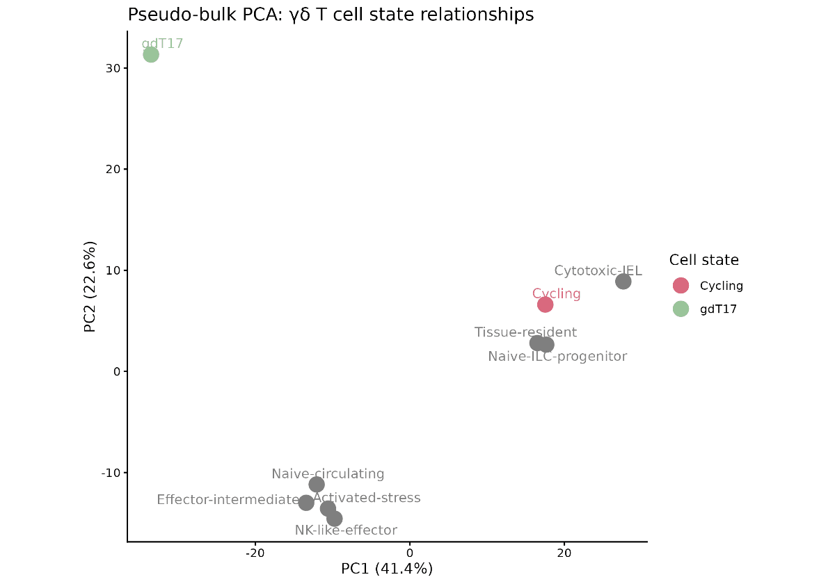
Transcriptional Structure of γδ T Cell States
Pseudo-bulk PCA, computed on aggregated expression profiles per cell type, revealed that gdT17 cells form a distinct transcriptional axis, separating maximally along PC1 from other γδ T cell states .
Effector and NK-like populations clustered more closely together, while naive and tissue-resident states occupied separate regions of transcriptional space, reflecting functional stratification.
Data Quality Considerations
HTO demultiplexing performance varied significantly across batches. The DEC batch showed sparse and unimodal HTO distributions, indicative of poor labeling quality, and was excluded to prevent misassignment of tissue identity .
As a result, tissue-resolved conclusions are derived from a single high-quality batch (OCT), and certain tissues (e.g., spleen, lymph node, skin) are not represented in the final analysis.
Translational Relevance
This analysis support downstream applications in:
Target discovery in immune-mediated diseases
Tissue-specific biomarker identification
Mechanistic profiling of immune cell states in therapeutic contexts