

This use case was co-created by academic researchers and Drylab. The full chat conversation demonstrates how researchers interact with Drylab to perform analyses, ask questions, and adapt workflows as new insights emerge.
We hope these examples inspire new approaches and possibilities in your own research.
Methods Summary
CZI image files from healthy and disease mouse liver tissue were processed using a Python-based pipeline (czifile package) to extract multi-channel z-stack data. Maximum intensity projections were generated across 20 z-slices to create 512×512 pixel composite images. Channel assignment was based on fluorophore emission wavelengths and visual inspection of staining patterns:
CD31 (AF488): Channel 1 - pan-endothelial marker showing extensive vascular network
LYVE1 (Cy3): Channel 2 - lymphatic endothelial marker with similar but sparser pattern
Target X (AF647): Channel 3 - diffuse cytoplasmic staining
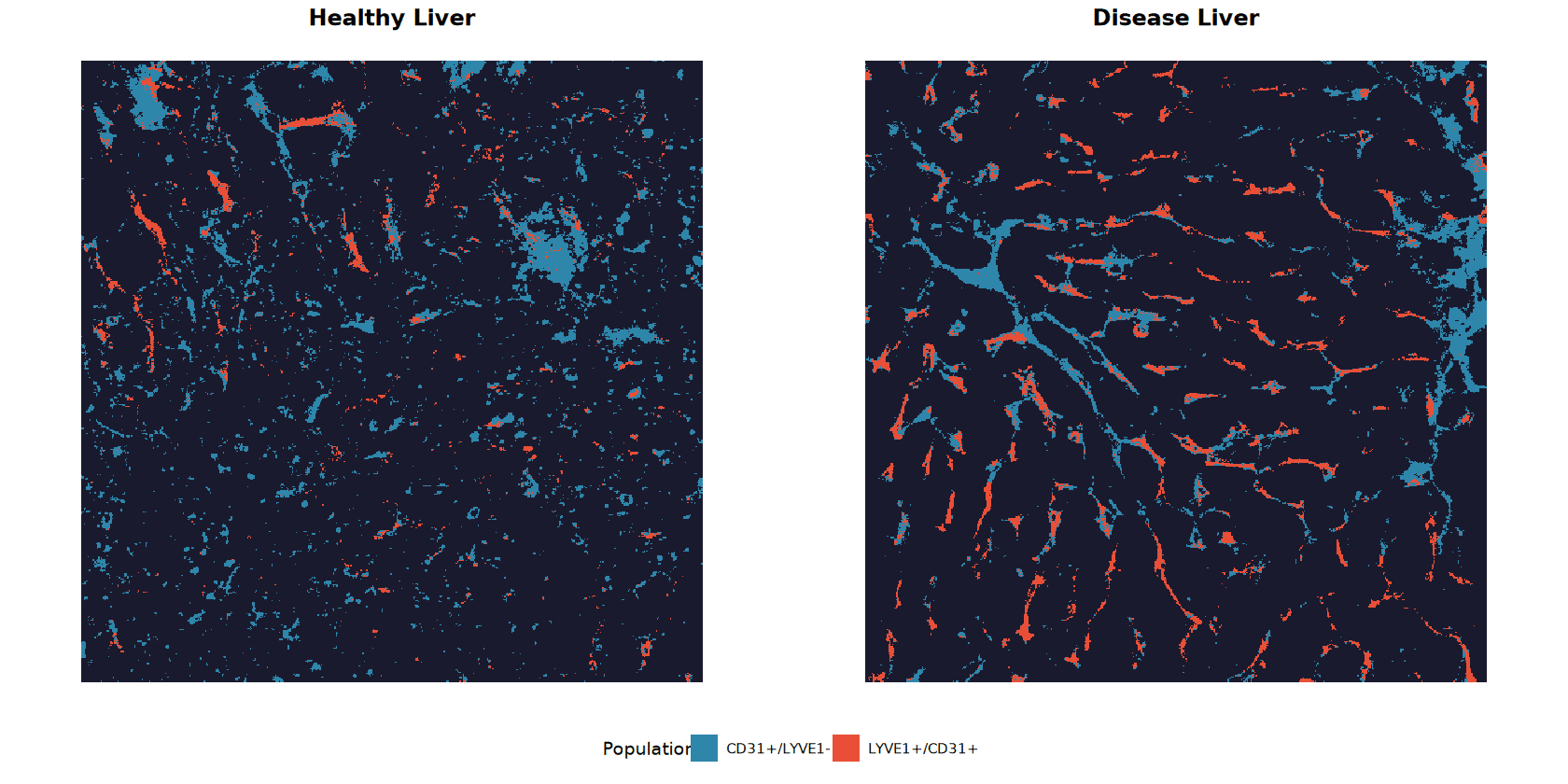
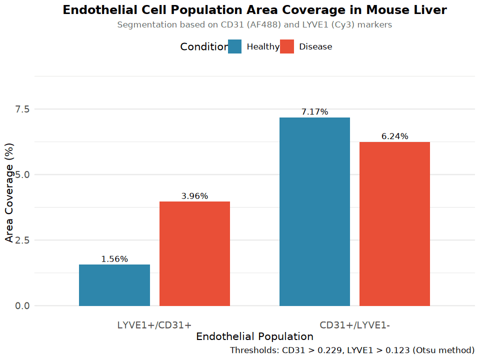
Segmentation was performed using Otsu's automatic thresholding method (CD31 threshold = 0.229, LYVE1 threshold = 0.123). Binary masks were combined to define two endothelial populations: LYVE1+/CD31+ (double-positive) and CD31+/LYVE1- (CD31 only).
Key Findings
Population | Healthy | Disease | Fold Change |
|---|---|---|---|
LYVE1+/CD31+ | 1.56% | 3.96% | 2.54x increase |
CD31+/LYVE1- | 7.17% | 6.24% | 0.87x (slight decrease) |
Total CD31+ | 8.73% | 10.19% | 1.17x |
Total LYVE1+ | 6.27% | 7.61% | 1.21x |
The quantification reveals a striking 2.54-fold expansion of LYVE1+/CD31+ double-positive endothelium in disease liver compared to healthy, while the CD31+/LYVE1- population shows a modest 13% decrease.
Interpretation
The segmentation overlay reveals marked morphological changes between conditions:
Key biological insights:
The preferential expansion of LYVE1+/CD31+ endothelium in disease liver is consistent with liver sinusoidal endothelial cell (LSEC) remodeling commonly observed in hepatic pathologies such as fibrosis or chronic inflammation
LYVE1 is a characteristic marker of LSECs and lymphatic vessels; its increased co-expression with CD31 suggests either LSEC proliferation/activation or lymphangiogenesis
The relative stability of the CD31+/LYVE1- compartment indicates that conventional blood vascular endothelium remains largely unchanged, while the sinusoidal/lymphatic compartment undergoes pathological expansion
Disease liver shows more elongated, interconnected vascular structures compared to the fragmented pattern in healthy tissue, indicative of vascular remodeling
Recommended Next Steps
Quantify multiple fields of view per condition to enable statistical testing (current analysis represents single images)
Include Target X (AF647) channel in the analysis to correlate its expression with endothelial populations
Consider adaptive thresholding or machine learning-based segmentation for improved accuracy
Perform morphometric analysis (vessel length, branching, diameter) to characterize structural changes
Validate findings with additional biological replicates (n≥3 per condition)
